Apoptosis vs. Necrosis: Disease Mechanisms Explained
Need help with this assignment?Get an original answer from a qualified tutor — from $10/page.
Get it written →Apoptosis vs necrosis is a core comparison in advanced pathophysiology. Both end in cell death, but they differ in triggers, mechanisms, appearance and consequences for surrounding tissue. This guide explains both and shows how they appear in disease.
Quick answer
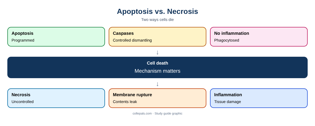
Apoptosis is regulated, energy-dependent programmed cell death of single cells, driven by caspases through intrinsic (mitochondrial) or extrinsic (death receptor) pathways; cells shrink and are removed without inflammation. Necrosis is uncontrolled death of groups of cells after severe injury; cells swell and rupture, releasing contents that trigger inflammation.
Examples and patient cases on this page are fictional and written by Collepals for study purposes. NURS 6501 is Walden University’s Advanced Pathophysiology course; assignments vary by term, so follow your own syllabus and rubric. This content is educational, not clinical advice. Collepals is not affiliated with Walden University.
Sample document: an apoptosis vs. necrosis comparison sheet (Word).
Apoptosis vs. Necrosis Compared
| Feature | Apoptosis | Necrosis |
|---|---|---|
| Trigger | Physiologic (development, turnover) or pathologic (DNA damage, viral infection, immune killing) | Severe injury: ischemia, toxins, infection |
| Cells affected | Single cells | Groups of cells, tissue |
| Cell size | Shrinkage | Swelling |
| Membrane | Intact; blebs form apoptotic bodies | Ruptured |
| Mechanism | Regulated: caspase activation via intrinsic (mitochondrial) or extrinsic (death receptor) pathways | Unregulated: ATP loss, enzyme digestion |
| Inflammation | None; phagocytosed quietly | Present; contents leak |
| Examples | Embryonic development, cancer cell elimination, cytotoxic T-cell killing | Myocardial infarction (coagulative), brain infarct (liquefactive), TB (caseous) |
Pathways of Apoptosis
- Intrinsic (mitochondrial): DNA damage or cell stress shifts the balance of BCL-2 family proteins, releasing cytochrome c and activating caspase-9.
- Extrinsic (death receptor): Fas ligand or TNF binds death receptors, activating caspase-8.
- Both converge on executioner caspases (such as caspase-3) that dismantle the cell (Rogers, 2023).
Types of Necrosis
- Coagulative: preserved tissue architecture; ischemic heart and kidney.
- Liquefactive: digested tissue; brain infarcts, abscesses.
- Caseous: cheese-like; tuberculosis.
- Fat necrosis: pancreatitis.
- Gangrenous: limb ischemia, often with infection.
Why It Matters Clinically
Too little apoptosis allows damaged cells to survive, contributing to cancer and autoimmunity (see Cancer: Cellular Mechanisms, Tumor Growth and Metastasis and Autoimmune Disease: Pathophysiology and Disease Progression). Too much contributes to neurodegeneration. Necrosis releases enzymes such as troponin from cardiac cells, which is why biomarkers rise after myocardial infarction.
Frequently Asked Questions
Does apoptosis cause inflammation?
No. Apoptotic bodies are phagocytosed without inflammation.
Which type of necrosis occurs in a stroke?
Liquefactive necrosis.
Why do cancer cells avoid apoptosis?
Mutations, such as loss of p53 or overexpression of BCL-2, block death signals.
Get Help Explaining Apoptosis and Necrosis
Advanced pathophysiology assignments demand precise mechanisms, current evidence and clear APA writing. Collepals.com helps NURS 6501 and other advanced pathophysiology students:
- Explain disease mechanisms step by step, from cells to symptoms.
- Write case study analyses, discussion posts and peer responses.
- Build concept maps, presentations and research papers.
- Find current evidence, cite in APA 7 and get drafts edited and proofread.
Browse our free model assignments or learn more about our nursing assignment writing service.
Use Collepals examples as study guides and follow your school’s academic integrity and AI policies.
More NURS 6501 and pathophysiology guides
Collepals is an independent study resource. Follow your program’s instructions and rubric and write your own work.
References
Hanahan, D. (2022). Hallmarks of cancer: New dimensions. Cancer Discovery, 12(1), 31–46. https://doi.org/10.1158/2159-8290.CD-21-1059
Rogers, J. L. (Ed.). (2023). McCance & Huether’s pathophysiology: The biologic basis for disease in adults and children (9th ed.). Elsevier.
Get a plagiarism-free answer to this question
Send us your instructions and we’ll match you with the best writer in your subject.
- 100% human-written, zero AI
- Turnitin report included
- Confidential — we never share your data
- Free revisions & refunds